


In today’s article we are going to talk about maxillary cysts, alesion that should be detected in its early stages to avoid its possible consequences. We will also explain how we solve it in the dental clinic.
What is a maxillary cyst and how is it detected?
A maxillary cyst is a benign lesion that appears in the upper jaw or mandible, in the form of a pocket and containing liquid, air or other material inside.
Maxillary cysts are usually painless if they are small and may go unnoticed. For this reason, we recommend periodic radiological analysis to detect any type of anomaly early and thus avoid its possible development.
In the
orthopantomography
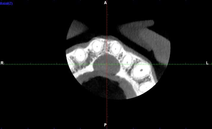
(2D radiograph of the whole mouth) the maxillary cyst is seen as a dark, rounded, pouch-like area. In case it is detected, it will be advisable to perform a CT scan (3D radiography) in order to appreciate the volume of the lesion and thus make an accurate diagnosis.
It should be taken into account that there are extensive maxillary cysts that can cause serious problems such as bone deformities, infections, dental mobility and rupture of the jaw in cases of giant cysts.
Why does a maxillary cyst appear?
Some cysts have a genetic cause. Among them are those related to the development of the jaws and teeth at early ages. For example, in children, if a tooth does not erupt correctly, it may be included within the jawbone, giving rise to a cyst.
Other cysts derive from deep caries that have reached the nerve. In these cases, an infection occurs inside the tooth that expands until it reaches the bone that supports it, forming a radicular cyst (type of cyst that comes from the tooth and is located at the tip of the root).
A maxillary cyst may also appear after trauma to the facial area, due to infections or due to a malfunction of the salivary glands.
Classification of maxillary cysts
Cysts are classified as odontogenic if they are related to dental structures (such as radicular, residual, paradental, follicular, eruption and keratocyst cysts) and non-odontogenic (such as naso-palatine, mid-palatine, globo-maxillary, naso-labial and mid-mandibular cysts).
The cover image (3D CT scan) shows a nasopalatine cyst, one of the most common cysts in the oral cavity. It can be seen as the black shadow behind the teeth.
How to fix a maxillary cyst?
If the cyst is related to the infection of the tooth, a root canal should be performed.
endodontics
and/or apical surgery to remove it:
This means removing all the bacteria from the tooth root, commonly called “killing the nerve” and/or removing the tip of the root next to the cyst (apical surgery). In the case of small cysts, endodontics alone will be sufficient to eliminate it (less invasive treatment), while there are other cases in which apical surgery or both interventions must be performed. Hence the importance of making a good diagnosis through 3D technology and specialists in endodontics and surgery to correctly and permanently eliminate the maxillary cyst.
In the case of extensive, fast-growing cysts or cysts that cause symptoms to the patient, the technique used is enucleation, which consists of eliminating the cyst in its entirety (together with the bag that encapsulates it) so that it does not reappear. Subsequently, the cyst is analyzed in the laboratory. During the procedure we use local anesthesia to make the procedure painless. In the post-operative period, the surgeon will provide you with a complete analgesic guideline and indications to avoid pain and promote the healing process.
Our multidisciplinary team will assess the type of cyst and decide the and decide which is the best treatment in each case and for each patient.