


In today’s article we are going to talk about orthopantomography, a radiological test necessary to be able to make an initial general diagnosis of a patient’s dental health. At the same time, we will also provide basic concepts on how to interpret it.
What is an orthopantomography?
Orthopantomography is a 2-dimensional frontal radiograph of the teeth and jaw bones of a human being. It is a dental technology that was created to provide the dentist with an overview of the internal structures of the teeth and the bone that supports them.
What is orthopantomography used for?
To make an initial general diagnosis of the oral health of any person, the dentist will perform an intraoral examination to check the external state of your teeth and gums.
Since this examination does not allow you to see the internal structure of the teeth and the bone that supports them, you will need to use a 2D panoramic X-ray, which we call orthopantomography, to complete the first general diagnosis.
With the intraoral exploration and the analysis of the orthopantomography, the professional will be able to identify if there are oral pathologies and if more specific 2 or 3 dimensional radiographs are necessary to complete a global and precise diagnosis.
What information can an orthopantomography give us?
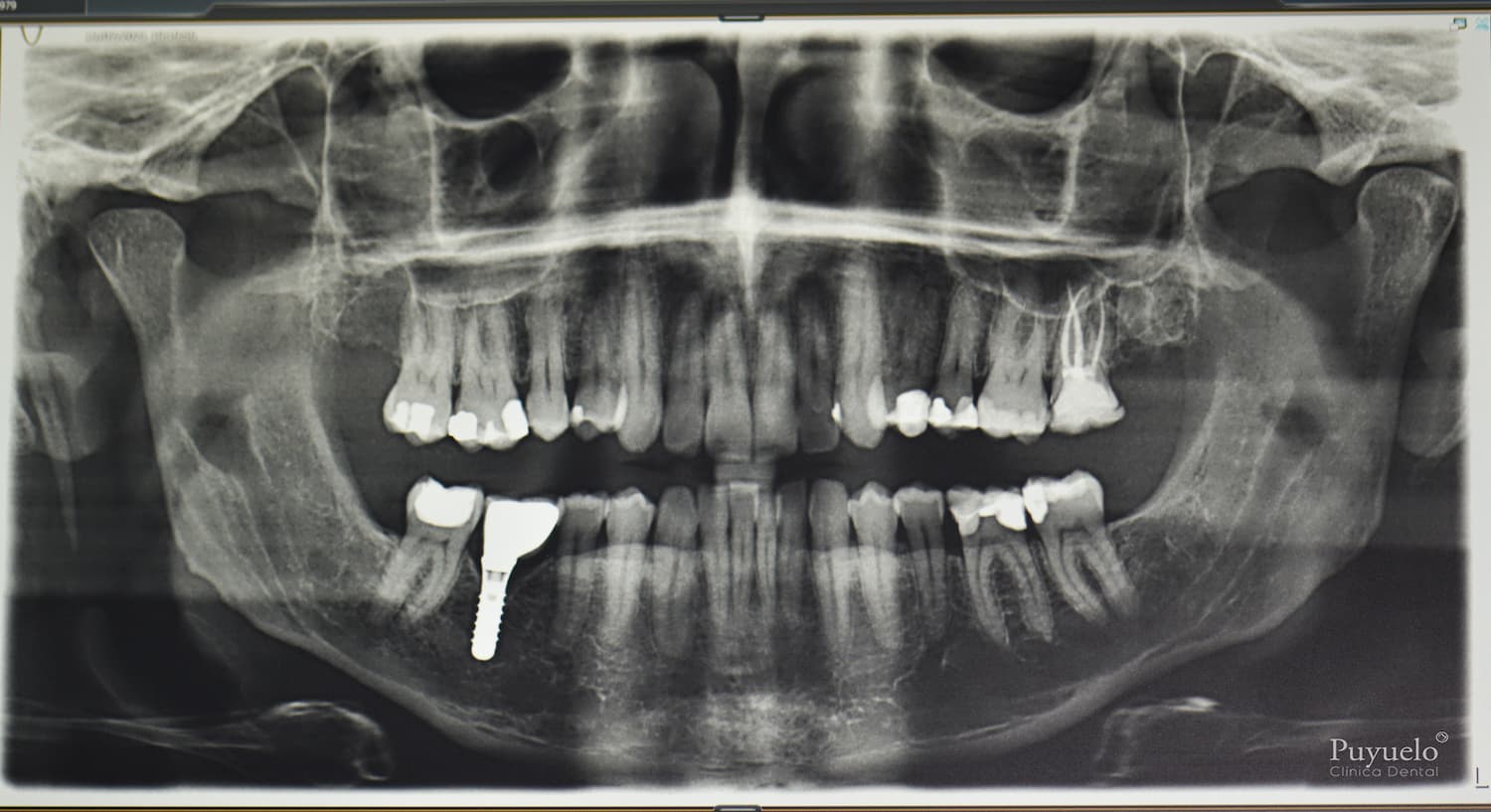
Orthopantomographies are seen in different shades between black and white, which we are going to classify:
- Gray tone: we can make out the patient’s teeth and bone. In a healthy mouth, 16 teeth will be observed in the upper arch and another 16 in the lower arch, as long as the wisdom teeth have not been extracted, which according to many articles do not serve any function and their extraction will be recommended if they compromise oral health.
The bone level is seen through a gray line that covers part of the teeth. A healthy patient will have the bone level just where the dental crown ends and the root begins.
If the bone line is below, it means that, either currently or at some period, there have been bacteria under the gum that have been deteriorating it. Hence the importance of visiting a dental clinic specializing in gums and dental prevention to slow bone loss.
- White shade: we can see the material of the treatments carried out in the mouth due to some pathology that has affected the tooth or due to esthetic rehabilitations.
Root canals are seen as two white sticks that fill the roots of the tooth, it will be important that they reach the apex to prevent bacteria from entering. Composite reconstructions or ceramic restorations are seen in the crown portion of teeth that have suffered decay or wear. Implants are seen as a screw that replaces the root of the tooth and is screwed to a ceramic crown.
- Black shade: Corresponds to everything that is not tooth, bone or any other material. If we observe the black color in the bone or any tooth, it will mean that there are bacteria that are deteriorating them. First of all, more specific 2D or 3D radiographs of the affected area should be taken to obtain the maximum information and to be able to make an accurate diagnosis. If the presence of bacteria is confirmed, action should be taken to remove the bacteria and reconstruct the affected tooth or tooth support (bone and/or gingiva).


